A new lens onthe blood film.
Smart-Eye clips onto the microscope your laboratory already owns and sorts routine films into three queues before anyone sits down at the bench. The model runs on the phone. Nothing leaves the room.
The microscope isn't the problem.
Attention is.
Light microscopy is still the reference standard for identifying parasite species and measuring density. But a busy laboratory runs it the way it did in 1990 — one scientist, one eyepiece, hundreds of slides, and no way to know which ones matter until they have all been read. Fatigue does the rest.
The real cost isn't time. It's that expert judgement gets spent confirming negatives instead of on the slides that genuinely need it.
Three parts. All of them
fit the lab you already have.
Smart-Eye does not ask anyone to replace a microscope. It adds a layer of attention on top of the one in the room.
The adapter
A machined cradle that clamps onto a standard eyepiece and aligns a phone camera to the optical path under a 100× oil objective. It bolts nothing, drills nothing, and comes off in seconds.
The capture app
Walks the technician through a fixed field-of-view protocol, holds exposure steady, and tags the sample. Everything is cached on the handset — there is no upload step to fail.
The engine
A vision model that segments cells, finds ring-forms and chromatin dots, and scores each film. Inference happens on the device, so latency is a second and patient images never leave the bench.
It reads the film first,
so a person doesn't have to read them all.
Capture
The technician prepares the film as always, mounts the phone, and shoots the guided sequence of fields. The app checks focus, contrast and stain quality as it goes and asks for a re-shoot when a field won't hold up.
8–10 minRead
On the handset, the model segments red and white cells to build a denominator, then scans for the intracellular morphology that distinguishes Plasmodium species. It returns a score, a density estimate and the regions it based them on.
1–2 minVerify
The film lands in one of three queues with its evidence attached. A scientist confirms, corrects, and signs. The report template is already filled in and exports over WhatsApp or into the LIS.
1–2 minEvery film ends up
in front of the right amount of attention.
Smart-Eye does not decide anything. It sorts. The scientist still verifies, interprets and signs every report — the platform only changes the order in which they arrive and how much is already known about each one.
Likely negative
No identifiable intracellular parasite across the standard set of digital fields.
Confirm quickly and sign. Nothing here needs a second opinion.
Suspicious
Ambiguous objects, low-density features, or morphology the model won't commit to.
Scan the flagged fields at concentration before drawing a conclusion.
High priority
Clear, high-density parasitaemia or a severe-indicator signature.
Goes to the top of the bench. Species confirmation, now.
Cycle times are operational targets for the first rollout, drawn from workflow observation in working laboratories. Sensitivity and specificity are being benchmarked against consensus readings from WHO-certified microscopists and PCR confirmation under IRB-supervised validation.
Fourteen days, free.
Then you look at the data.
We would rather a laboratory judge this on its own floor than on a slide deck. Every prospective partner gets a structured two-week validation pilot at zero upfront cost, and the evidence it produces belongs to them.
We come and look
Daily microscopy volume, where the queue actually backs up, who is on which bench. No hardware arrives until we understand the lab.
Installed at no cost
The founder fits and calibrates the adapter to your microscope and your light source personally. Nothing to pay, nothing to sign.
Side by side
Your technicians run their normal malaria requests with Smart-Eye alongside. We watch, support and record. The lab runs as it always does.
Your numbers, not ours
An audit built from your own data: turnaround against your manual baseline, scientist-hours recovered per shift, repeat reviews avoided — then you decide, either way.
Smart-Eye is a clinical decision-support tool. Every result requires verification, interpretation and formal sign-off by a registered Medical Laboratory Scientist — during the pilot and after it.
No lab should have to throw out its microscope to join the digital era.
Smart-Eye was built by a Medical Laboratory Scientist who has stood at the bench it is meant to fix. The models are trained on the stain variation, cell density and artefact noise of West African laboratories specifically — the things a model trained elsewhere quietly gets wrong.
Built by a scientist.
Checked by practitioners.
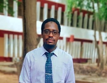
Medical Laboratory Scientist, Ahmadu Bello University, Zaria. Worked in histopathology, haematology and clinical chemistry, with research in automated digital cell quantification.
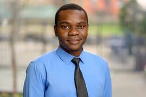
Computational oncologist. Research fellow at Memorial Sloan Kettering; PhD in molecular pathology, Tokyo University of Science; senior lecturer at Ahmadu Bello University.

Immunologist, 13 years lecturing at Ahmadu Bello University and honorary Medical Laboratory Scientist at ABUTH, Zaria. Gates Foundation and NIH/FIC awardee.
Put it on your bench
for two weeks.
For laboratory managers and medical directors. Tell us what your microscopy load looks like and we'll come and see it. We reply within five working days.
Or write to us directly at info@smarteyediagnostics.com